




What is a neuron
A neuron is a specialized cell in the nervous system that receives and sends messages within that system. It is the building block of nervous system.
The human brain has 100 billion neurons, each neuron connected to 10 thousand other neurons. Sitting on your shoulders is the most complicated object in the known universe. — Michio Kaku
Functions of a neuron
- Receiving signals
- Processing information
- Conducting impulses
- Transmitting signals to other cells
- Communication and control of bodily functions
Structure of a neuron

- Dendrite-These are branch like structures that receives messages from other neurons.
- Soma – The central part of the neuron that contains the nucleus and cell organelles.
- Axon– A long, thin structure that carries electrical impulses from the soma to the axon terminals.
- Axon hillock– The point where axon begins.
- Myelin sheath – Axons are covered with a fatty layer called myelin sheath. It protects the axon as well as speed up the transmission of nerve impulses. Oligodendrocytes produce myelin sheath for the Central nervous system and Shwann cells produce myelin sheath for the peripheral nervous system.
- Nodes of ranvier– The gaps in the myelin sheath along the axon.
- Axon terminals– Located at the end of axons, axon terminals are responsible for transmitting signals to other neurons.
- Synapse– At the end of axon terminals, there is a gap between two neurons called synapse. Neurotransmitters are released from terminal buttons to the synapse and carry signals across other neurons..
Types of neurons
Based on function:-
- Afferent neurons- Carry information from sensory receptors to the Central nervous system (CNS).
- Efferent neurons- Transmits signals from the CNS to muscles or glands to produce a response
- Interneuron- Connect sensory and motor neurons within the CNS and process information.
Based on structure :-
- Unipolar neurons- Have only one axon.
- Bipolar neurons- These neurons have one axon and one dendrite
- Multipolar neurons- These neurons have a single axon and multiple dendrites.
Glial cells
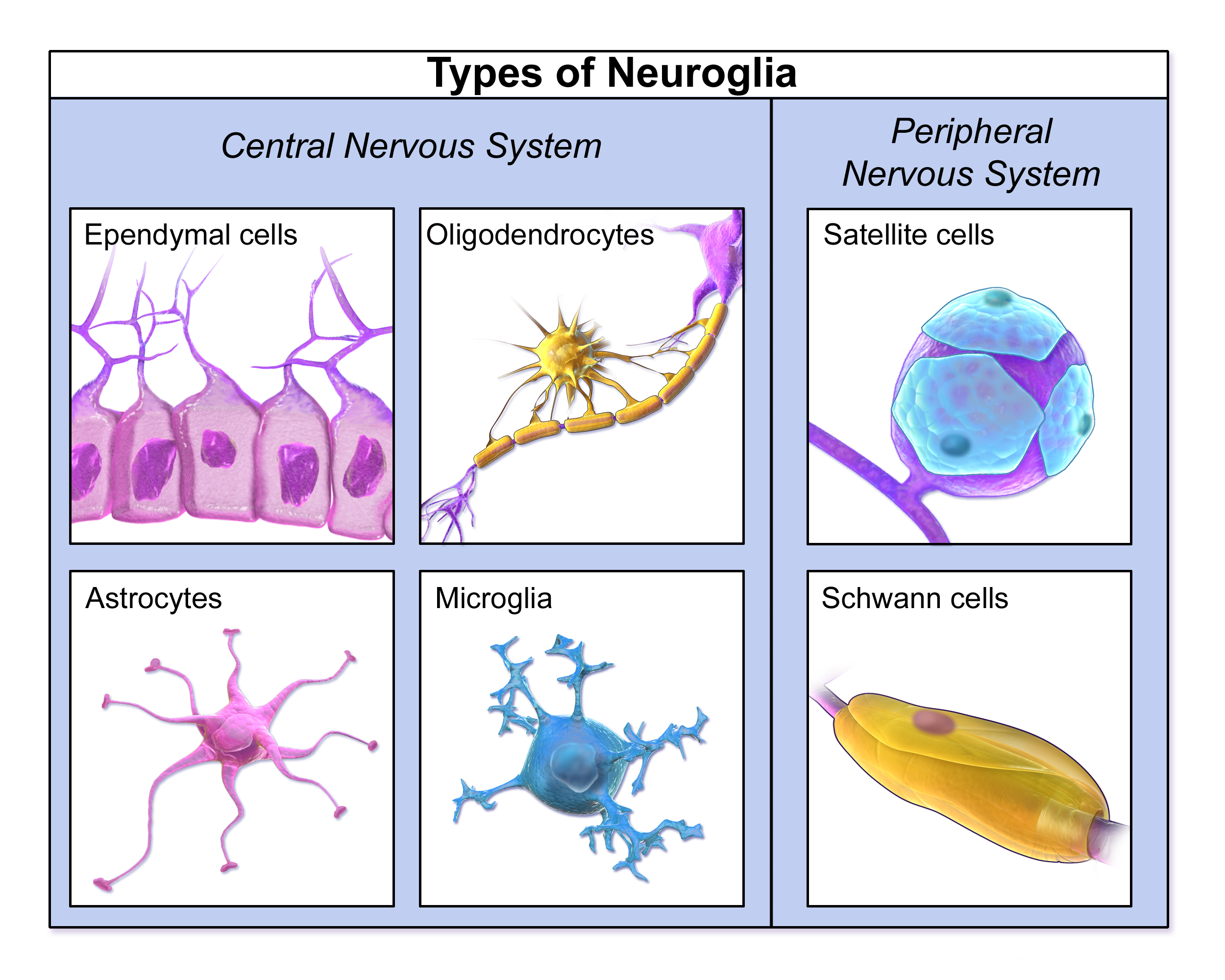
Glial cells, also known as neuroglia are an integral part of the human nervous system. The functions of glial cells include helping to support and nourish nerve cells, or neurons. Glial cells are not actually a type of neuron but similar to neurons Glial cells are also found in Central nervous system as well as Peripheral nervous system.
| Found in the CNS :- | Found in the PNS:- |
| 1. Microglia- Remove debris, pathogens and damaged cells through Phagocytosis. 2. Ependymal cells- Produces cerebrospinal fluid (CSF) 3. Oligodendrocytes- Produces myelin sheath for the CNS 4. Astrocytes- Maintains Blood Brain Barrier (BBB) | 1. Satellite cells – Provide support and nutrients to neurons 2. Shwann cells- Produces myelin sheath for the PNS. |
Neural Impulse
- Resting potential– At rest, a neuron has more negative charge inside and more positive charge outside the cell. This charge difference is due to the unequal distribution of ions maintained by both diffusion (movement from high to low concentration) and electrostatic pressure (attraction between opposite charges). Inside the cell are positively charged potassium ions (K⁺) and large negatively charged proteins that can’t leave, keeping the inside negative. Outside, there are sodium ions (Na⁺) and chloride ions (Cl⁻), but sodium can’t enter because its channels are closed. The sodium ions gather near the membrane, attracted to the negative charge inside. This difference creates electrical potential preparing the neuron to fire when stimulated.
- Depolarization– When a neuron receives a strong enough stimulus, the neuron becomes depolarized. The inside of the neuron starts to become less negative typically reaching its threshold (-55 mV). The voltage gated sodium channels open allowing sodium ions to move inside.
- Repolarization– Once the neuron reaches its peak action potential (+40 mV), the sodium channels close and the potassium channels open, potassium ions move out of the neuron causing the inside of the cell to become negative again.
- Hyperpolarization– Sometimes the outflow of potassium overshoots and the membrane potential becomes more negative than the resting potential. This is the brief phase of hyperpolarization where neuron returns to its resting potential.
Action potential and Refractory period
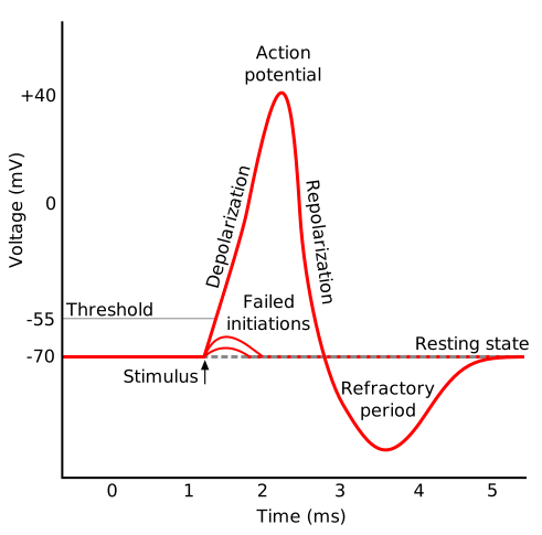
An action potential/ neural impulse is a rapid, temporary change in the electrical charge of a neuron’s membrane that allows it to transmit messages. The primary rule that governs action potentials is known as the All-or-None Law. This states that once the threshold level is reached neuron will fire at full strength—and if the threshold is not reached, no action potential will occur at all.
No Gradation: A stronger stimulus does not produce a stronger action potential; it may only increase the frequency of firing.
Threshold: The minimum level of stimulation (typically around -55 mV) required to trigger an action potential.
Following an action potential, the neuron enters a refractory period, a brief time during which it cannot fire another action potential. This period is divided into two phases:
- Absolute Refractory Period: No new action potential can be initiated, no matter how strong the stimulus.
- Relative Refractory Period: A stronger-than-usual stimulus may initiate another action potential
Communication between neurons

Presynpatic and postsynaptic neurons
The presynaptic neuron contains vesicles filled with neurotransmitters. The postsynaptic neuron’s dendrite contains ion channels with receptor sites that accept only specific neurotransmitter molecules—like a key fitting into a keyhole.
Release of neurotransmitters
When an action potential travels down the axon and reaches the axon terminal, it activates the synaptic vesicles. The vesicles release neurotransmitters into the synaptic gap. The neurotransmitter molecules float across the synapse toward the postsynaptic neuron.
Binding to receptor sites
These molecules fit into specific receptor sites on the postsynaptic ion channels, triggering a response. Neurotransmitters can either be Excitatory (making the postsynaptic neuron more likely to fire) or Inhibitory (making it less likely to fire).The effect depends on the type of neurotransmitter and the specific synapse.

Removal of neurotransmitters
After neurotransmitters have carried the message across the synaptic gap, they must be quickly removed to prevent continuous stimulation of the postsynaptic neuron.This is done through three main processes, Reuptake, Enzymatic degradation and Diffusion.
- Reuptake is the process by which neurotransmitters are reabsorbed back into the presynaptic neuron.
- Enzymatic degradation is the process where enzymes in the synaptic gap break down neurotransmitter molecules into inactive components.
- Diffusion- Some neurotransmitter molecules simply drift away from the synaptic gap and move into the surrounding.


Share this article by Power Within Psychology (PWP):

Discover more from Power Within Psychology | BLOGS
Subscribe to get the latest posts sent to your email.